Record
A provider-worn spatial device captures gait, tapping, tremor, and clinician-guided motor maneuvers during routine assessment.
Highlighted research · Spatial motor phenotyping
Parkinson’s disease is expressed through movement, yet the movement itself is rarely preserved. We test whether provider-worn spatial technology can capture the clinical motor exam and make change measurable across visits.
Read the rationale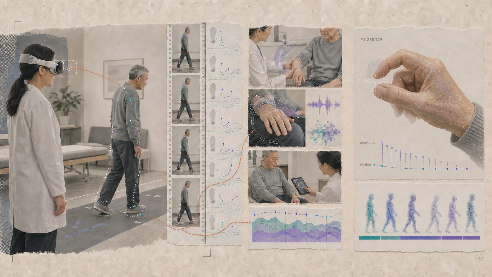
Rationale
Motor examinations translate gait, tremor, bradykinesia, finger tapping, and clinician-assessed rigidity into ordinal ratings. Those ratings are clinically essential, but they cannot retain the full kinematics of the encounter or its variation with medication and stimulation. The full kinematics are there in the exam; the score just can't hold them.
Provider-worn spatial video can preserve the motor exam, and machine-learned kinematics can track severity, fluctuation, and treatment response.
What we’re building
A spatial measurement framework built into the neurologic examination itself.
A provider-worn spatial device captures gait, tapping, tremor, and clinician-guided motor maneuvers during routine assessment.
Three-dimensional pose and hand kinematics describe step length, drag, asymmetry, amplitude, rhythm, decrement, and tremor.
Repeated visits are aligned to reveal patient-specific change across medication states, DBS settings, and disease progression.
The neurologist's exam stays at the center. The system adds objective kinematics and makes comparison across visits more precise.