Capture
A guided phone workflow helps acquire a usable image and flags problems with framing, distance, or image quality.
Highlighted research · Clinical imaging AI
Pressure injuries change over time, but their record is often reduced to a categorical stage and an inconsistent image. The question is whether routine clinical capture can yield a reproducible measure of wound state.
Read the rationale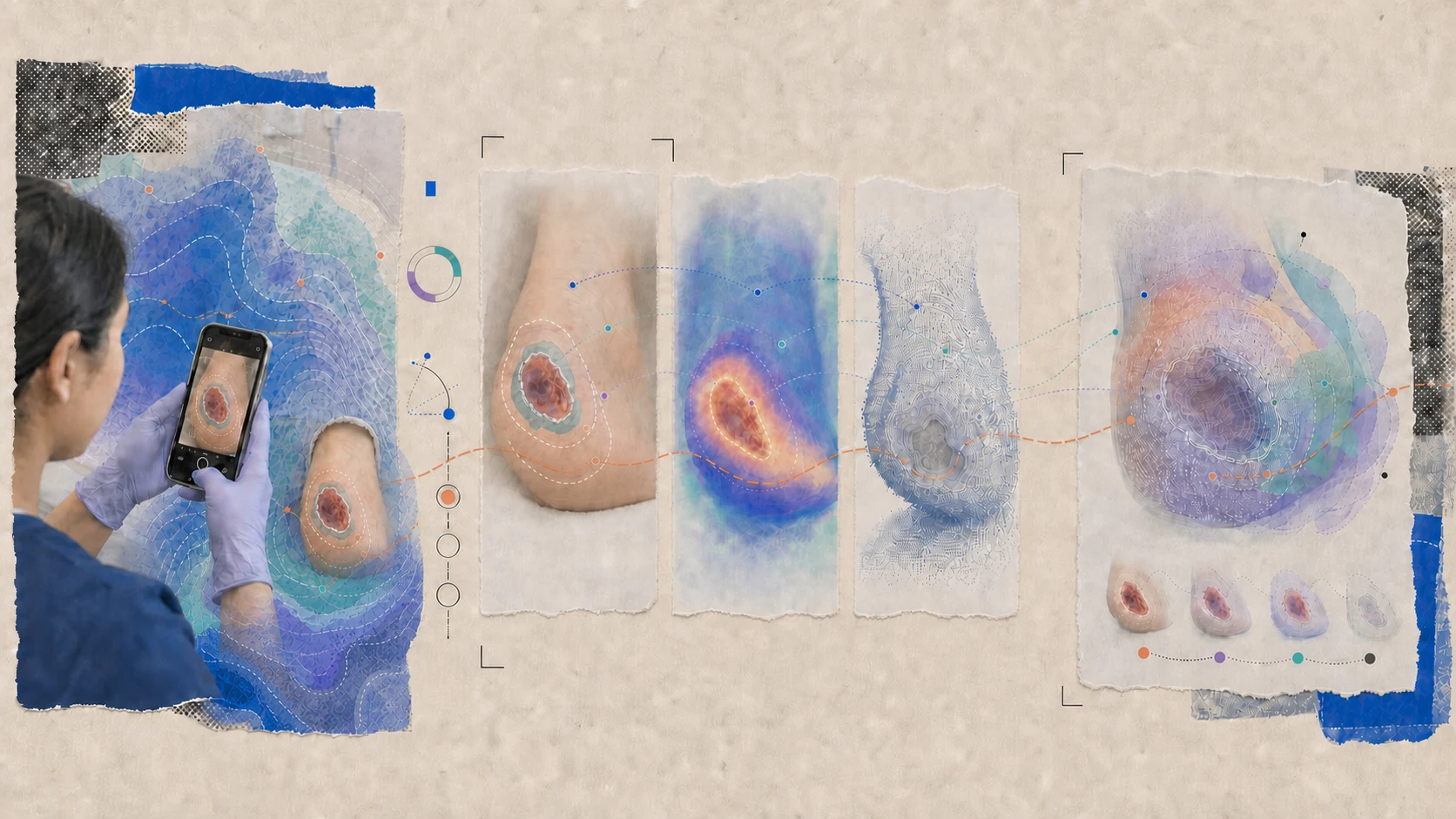
Rationale
Real photographs vary in lighting, distance, framing, device, wound presentation, and surrounding skin. Models trained only on small, curated image sets may perform well in familiar data while failing in the clinical environment where they are needed. Models have to be trained where care actually happens.
Combining real-world images with depth, infrared, and clinical context can produce a reliable digital biomarker of wound extent, tissue, and change.
What we’re building
A bedside workflow designed around the nurse and the patient—not around a curated research image.
A guided phone workflow helps acquire a usable image and flags problems with framing, distance, or image quality.
Image, spatial, thermal, and clinical signals are combined to characterize wound extent, tissue, depth, and change.
The system returns staging support and longitudinal comparison, with every output open to clinician review.
Staging decisions stay with clinicians. The system contributes a consistent measurement they can inspect, compare over time, and use at the bedside.