Acquire
A routine 12-lead ECG preserves synchronized electrical activity from twelve simultaneous viewpoints.
Highlighted research · ECG-AI
The 12-lead ECG is inexpensive, ubiquitous, and repeatedly collected across care. We search its waveform for latent signatures of ventricular function that conventional interpretation does not routinely expose.
Read the rationale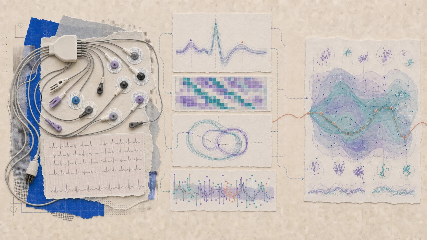
Rationale
A standard ECG records electrical activity from multiple anatomical viewpoints, but conventional interpretation focuses on established visible patterns. Echocardiography provides more detailed structural and functional information, yet it is less frequently obtained and more resource intensive. Subtle physiology may already be distributed across the waveform.
Deep learning across paired ECG and echocardiography data can identify distributed waveform signatures of ventricular dysfunction and create scalable cardiac screening biomarkers.
What we’re building
A predictive framework that learns from routine signals already embedded throughout clinical care.
A routine 12-lead ECG preserves synchronized electrical activity from twelve simultaneous viewpoints.
Waveforms are time-aligned with echocardiography, clinical context, and outcomes that define cardiac structure and function.
Models learn waveform signatures that can identify patients who may benefit from confirmatory imaging or closer evaluation.
The aim is a widely available test that surfaces hidden physiology and helps target echocardiography where it's needed most.